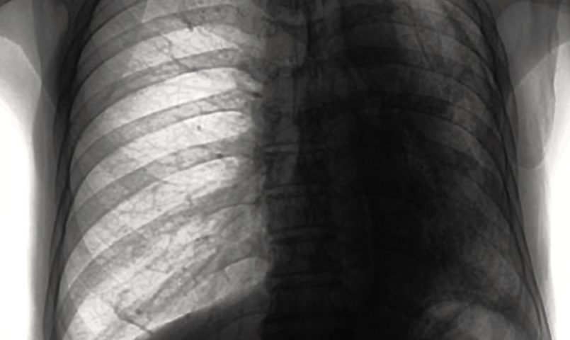
A pleural biopsy is a more accurate and efficient method of diagnosing malignant diseases that cause fluid leakage in the lungs than pleural effusion cell block (a cell preparation using pleural fluid), according to a new study.
The study, “Diagnostic Utility Of Pleural Fluid Cell Block Versus Pleural Biopsy Collected By Flex-Rigid Pleuroscopy For Malignant Pleural Disease: A Single Center Retrospective Analysis,” was published in the journal PLoS One.
The current methods used to diagnose pleural diseases with pleural effusion (accumulation of fluid between the two membranes that surround the lungs), such as malignant mesotholioma, include pleural biopsy and pleural effusion cell block.
The two methods have different approaches when preparing the samples for analysis: While pleural biopsy consists of spinning the cells collected with the biopsy directly into a glass slide, pleural effusion cell block includes the cells in paraffin blocks, which are later cut into thin layers for analysis. The latter approach better preserves the morphology of the collected cells, but until now, no studies had compared the diagnostic utility of both methods.
To address this question, researchers analyzed medical data from 68 patients who had undergone pleural effusion cell block and pleural biopsy through flex-rigid pleuroscopy (an analysis of the pleura using less rigid instruments to collect fluid) for malignant pleural disease with pleural effusion treated at the National Cancer Center Hospital in Japan between April 2011 and June 2014.
During the procedures, a minimum of 150 ml of pleural fluid was collected by pleuroscopy, followed by pleural biopsies from the abnormal site.
From the original group of patients, 35 were diagnosed with malignant pleural disease, such as adenocarcinoma (24 patients), combined adeno-small cell carcinoma (one patient), malignant pleural mesothelioma (MPM, seven patients), and metastatic breast cancer (three patients).
The aid to diagnosis was significantly higher using pleural biopsy than by cell block (94.2% vs. 71.4%). Indeed, all patients with a positive result on cell block also showed a positive result on pleural biopsy, but eight patients that showed negative results on cell block had positive results on pleural biopsy.
Two patients with negative results in both exams were diagnosed with sarcomatoid MPM by computed tomography scan-guided biopsy and epithelioid MPM during autopsy.
Together, these results suggest that pleural biopsy was more accurate in the diagnosis of pleural diseases.
The authors reported that their study has several limitations, such as the small sample size, and the fact that all patients came from the same cancer center, which can add some biases to the analysis.
“Pleural biopsy … was efficient in the diagnosis of malignant pleural diseases,” the researchers wrote. “Flex-rigid pleuroscopy with pleural biopsy and pleural effusion cell block analysis should be considered as the initial diagnostic approach for malignant pleural diseases presenting with [pleural] effusion.”


