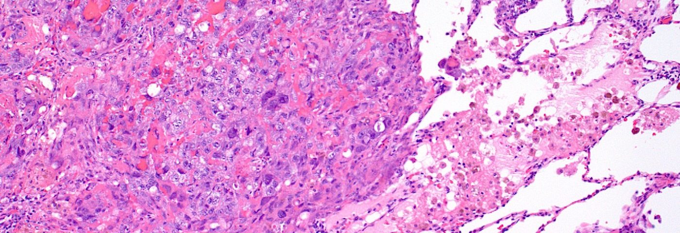
German researchers have come up with a new tissue-sample imaging and collecting technique for identifying diffuse malignant mesothelioma (DMM) subtypes.
The method that the team at Ruhr-University Bochum developed is based on advanced imaging and micro-dissection. It provides a more accurate molecular picture of DMM tissue samples and better identification of tissue biomarkers, they said. Micro-dissection involves using a microscope to dissect tissue.
Scientific Reports published the study, titled “Spatial and molecular resolution of diffuse malignant mesothelioma heterogeneity by integrating label-free FTIR imaging, laser capture microdissection and proteomics.”
“Diffuse malignant mesothelioma (DMM) is a heterogeneous malignant neoplasia manifesting with three subtypes: epithelioid, sarcomatoid and biphasic,” the researchers wrote.
DMM diagnosis usually relies on tumor characteristics, including its structure and its expression of specific biomarkers.
But DMM is similar to sarcomas and adenocarcinoma of the lung, which means doctors often have a hard time distinguishing the diseases and correctly diagnosing DMM. Making it even harder is that there are only slight differences between the DMM subtypes: epithelioid, sarcomatoid, and biphasic.
That means it’s crucial for doctors to identify biomarkers that can help them make an accurate diagnosis of DMM and its subtypes.
Researchers used Fourier transform infrared (FTIR) imaging guided tissue annotation instead of standard tissue staining to identify subtypes of DMM.
When a thin tissue sample is illuminated, it absorbs and reflects different amounts of light, depending on its structure and composition. This means scientists can use FTIR imaging to find a tissue’s molecular and structural fingerprint. And from that footprint they can identify the DMM subtype it belongs to.
“The robust and objective results produced by FTIR imaging minimized inter- and intra-observer variability and allow for automated, spatially resolved label-free annotation of tissue samples,” the researchers wrote.
The scientists used FTIR analysis to identify tumor subtypes in 17 tissue samples taken from 14 DMM patients. FTIR was more accurate than current methods of identification, the researchers said.
Coupling the imaging with laser-based micro-dissection allowed the team to collect micro-scale tissue samples without picking up adjacent tissue that could contaminate the results.
The combination also could lead to more accurate readings of protein levels, the team said. And that, in turn, could lead to new biomarkers for both DMM and its subtypes.
“The novel FTIR-guided LCM approach, combined with high-resolution proteome analysis, paves the way to more specific and predictive protein biomarkers,” the team wrote.


